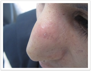
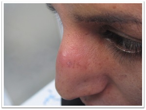
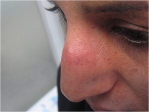
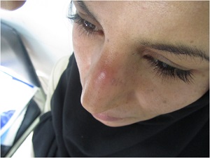
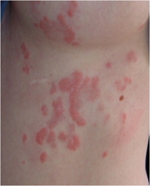
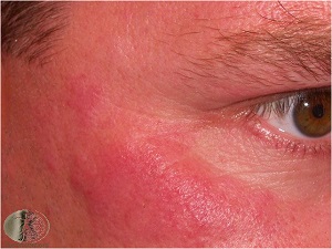
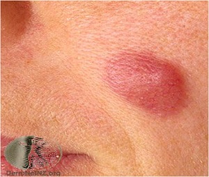
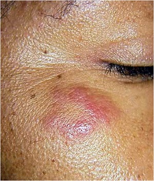
بیمار خانمی 31 ساله که از حدود 9 ماه قبل دچار یک پلاک ایندوره،
روی بینی شده است که بدون درد ولی با سوزش وخارش شدید همراه بوده
است.
در معاینه به جز ضایعه پوستی مشکل دیگری ندارد.
علایم سیستمیک ندارد و آزمایشات روتین بیماز نرمال است.
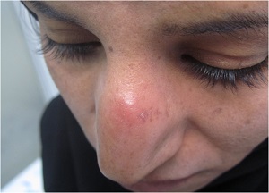
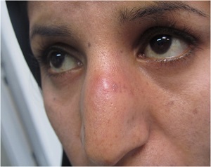
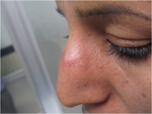
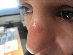
What is your diagnosis?
Lymphocytic infiltrate of Jessner
Epidemiology
-Lymphocytic infiltrate of Jessner occurs with equal incidence in men and women, and it is a disease primarily of middle-aged adults.
-It is very rare in children.
-Various authors believe it is a variant of either lupus erythematosus, cutaneous lymphoid hyperplasia or polymorphous light eruption.
-Others believe it to represent an infectious process
possiblyrelated to Borrelia burgdorferi infection
-There are cases of co-occurrence with
lupus erythematosus and with polymorphous light eruption
-Rare cases of drug-induced lymphocytic infiltrate of Jessner have been described.
-glatiramer acetate,
angiotensin-converting enzyme (ACE) inhibitors
Clinical features
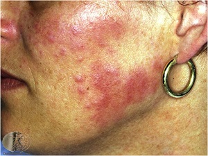
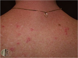
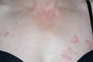
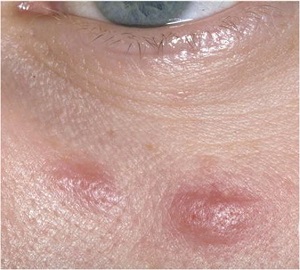
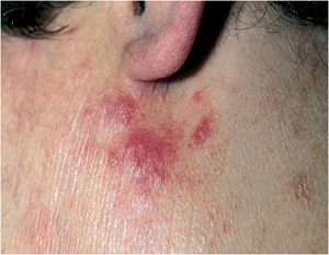
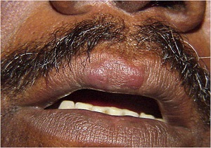
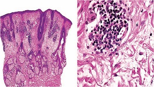
-Lymphocytic infiltrate of Jessner most commonly appears on the head, neck and upper back as one or several asymptomatic erythematous papules, plaques and, less commonly, nodules
-There are no secondary changes, such as scale, and annular plaques with central clearing are commonly observed.
- There are no systemic manifestations associated with lymphocytic infiltrate of Jessner.
.
-The eruption resolves spontaneously and without sequelae in most patients
Differential diagnosis
the plaque form of polymorphous
light eruption
cutaneous lymphoid hyperplasia
Cutaneous lymphoma
lupus erythematosus
�� Treatment
The cutaneous manifestations of lymphocytic infiltrate of Jessner resolve spontaneously within months to years, and they do not result in scarring.
Oral antibiotics and topical or intralesional corticosteroids
have been used with limited success.
Up to 50% of patients may improve with hydroxychloroquine.
Lymphocytic infiltrate of Jessner is generally resistant to radiation therapy
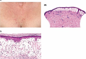
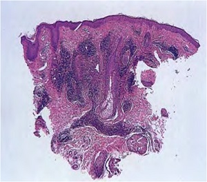
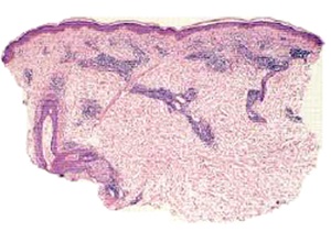
Pathology
Dense lymphocytic perivascular infiltrate, superficial and deep.
Dermal mucin is usually increased.
Epidermis is normal (unlike in lupus erythematosus
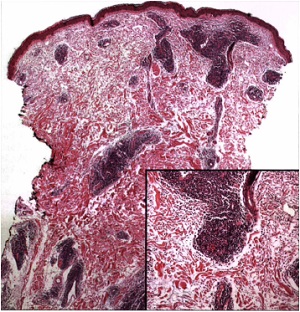
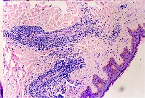
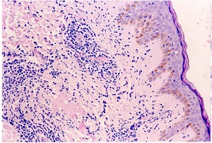
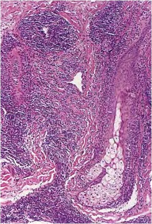
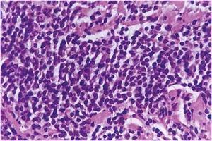
Superficial and deep perivascular and periadnexal dermatitis
Infiltrates of lymphocytes are accompanied by mucin in abundance in the interstitium.
Superficial and deep perivascular inflammation�8Ls+ DRUGS
8Ls :
Light reaction(Photocontact allergic dermatosis, polymorphous light eruption)
Lymphoma (SLL/CLL , B-cell type CD20+ , CD5+)
Leprosy
Lues(Syphilis)
Lichen striatus
LE (Tumid lupus and DLE)
Lipoidica necrobiosis
Lepidoptera(arthropods bite)
DRUGS:
Drug reaction and Dermatophyte
Reticular erythematous mucinosis
Urticarial stage of bullous pemphigoid
Gyrate erythema
Scleroderma,( localized variant)
B-cells in Jessner vs. T-cells in Lupus
DIF negative (in 10-20% of lupus cases DIF is negative)
No vacuolar changes
No epidermal atrophy
No follicular plug
Mucin may be seen.
-Slight epidermal atrophy and focal thickening of the dermoepidermal junction more common in TL(tumid lupu)
-Lymphocytic infiltrate was less dense in TL than in Jessner.
Polymorphic light eruption :
Subepidermal edema
Eosinophils and afew neutrophils are sometimes present
No dermal mucin
No lichenoid reaction
Some basal vacuolation may be seen.
No basement membrane thickening