آقاي ساسان سلام
اگر بيماري در حال پيشرفت باشد،نقاط جديد بدن كه تازه دارند رنگدانه از دست مي دهند ولي هنوز روند پيشرفت ادامه دارد،خيلي سفيد نيستند،در خصوص سوال دوم،چنين آمپول و درماني وجود ندارد،بهترين درمان براي شما نور درماني هست .
As a pediatrician one of the most common presenting complaints is that of an infant with a rash and with the large number of possible diagnoses (on the order of more than 30!)
It is essential to be able to recognize the characteristic appearance of common lesions.
Furthermore, it is imperative to be able to identify life-threatening disease processes from benign, self-limiting disease.
Colour changes
Skin redness
Jaundice
Vascular tone
Harlequin colour changes
Cutis marmorata (6.7% **)
• Genital hyperpigmentation (5% Caucasians; 20% Mongolians)
• Desquamation (65%; 14.6% *)
• Sebaceous gland hyperplasia (48%; 31.8% †)
• Acne (1.2% ‡)
• Epstein pearls (56%; 88.7% †), Bohn pearls
• Milia (48%; 34.9% †; 19% *)
• Adnexal polyp of neonatal skin around nipple (4.1% §)
• Toxic erythema (35%; 40.8% §; 20.6% †; 15.7% *)
• Transient pustular melanosis (1.7% ‡)
• Miliaria crystallina (3.1%; 4% §; 14.6% †; 16.9% *)
• Miliaria rubra (4% §; 5.5% *)
• Sucking blisters (9.8%)
• Perianal dermatitis (18.9% §; 7% *)
Postmaturity in the neonate, and sometimes dysmaturity
Dif Dx:
Collodion baby syndrome
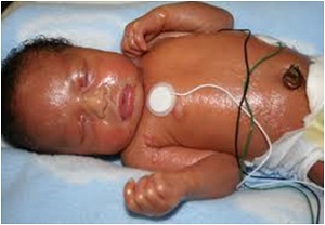
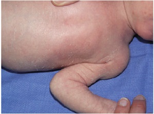
Harlequin colour changes
Harlequin colour change (HCC) is defined as transient erythema involving one - half of the infant ’ s body with simultaneous blanching of the other side and a sharp demarcation on the midline.
In some cases, HCC may be associated with tonic attacks and bradycardia, triggered by various factors, including defecation, rectal pain, perineal stimulation or emotion.
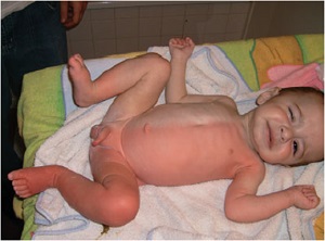
Aetiology.
Since original reports concerned mostly premature/low - weight babies or infants with intracranial injury, it was long thought that the abnormality in the central nervous control of the vascular tone was located at the hypothalamic level.
Indeed, most affected infants are in good general health with no other signs of dysregulation of vital functions.
Premature infants are more commonly affected than full - term infants.
Harlequin colour change usually occurs on the third or fourth day of life, though it has been noted up until the 21st day of life.
The midline demarcation of the erythema and the transitory colour changes (30 s to
20 min) are diagnostic
The face and genitalia are usually spared. The tongue and lips are unaffected.
Accentuation occurs following gravitational changes.
Turning the infant on the other side may induce blanchingof the red side and reddening of the pale side
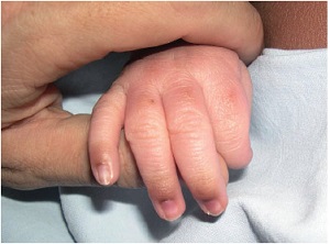
Cutis marmurata
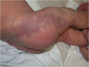
The genital area, and particularly the scrotum in male infants, and the dorsal surfaces of the distal phalanges of both hands and feet are the most frequently involved
Less frequently, nipple and areola, helix, umbilicus, and sometimes large skin folds
During the 1st yr
Infections
• Bacterial pustules: bullous impetigo, systemic sepsis (listerosis)
• Congenital cutaneous candidiasis
• Herpes, varicella, cytomegalovirus infection
Non-infectious disorders
• Incontinentia pigmenti
• Eosinophilic pustulosis (eosinophilic pustular folliculitis) of the scalp
• Infantile acropustulosis
• Omenn type of severe combined immunodefi ciency
• Buckley–Job hyperimmunoglobulin syndrome
• Self-healing histiocytosis
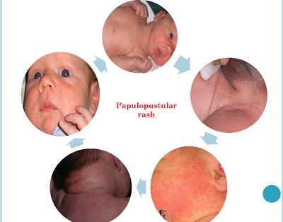
Sterile transient neonatal papulopustular eruptions
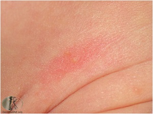
Erythema toxicum neonatorum (ETN)
Transient pustular melanosis (TPM)
Erythema toxicum neonatarum
is the most common transient rash in healthy term neonates, affecting 30-70 % of the newborns
First and forth days of life and lasts 2 or 3 days
The cause of ETN is not yet established
no predilection according to sex
White > Black
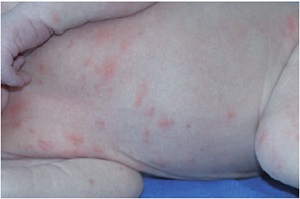
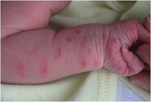
Lesions are located mostly on the trunk, with a predilection for the back, but are readily found on the upper arms, thighs and face
The palms and soles are relatively spared.
Flea bite picture (70%)
30% pustular
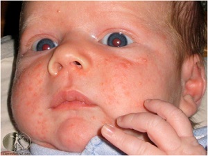
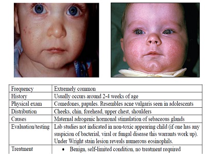
Neonatal acne
neonatal cephalic pustulosis is related to malassezia colonisation
Miliaria
The term miliaria is used to describe a group of transient eccrine disorders.
Occlusion of sweat ducts at various levels, resulting in leakage of sweat in the epidermis or papillary dermis
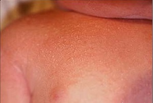
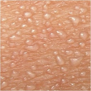
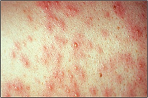
Milia
Milia are noted in 30 – 50% of neonates. They consist of 1 – 2 mm white or yellow papules on the nose, chin, cheeks and forehead.
The nose is predominantly affected. Less commonly, lesions may occur on the trunk and extremities.
Milia are epidermal cysts derived from the pilosebaceous follicles
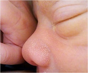
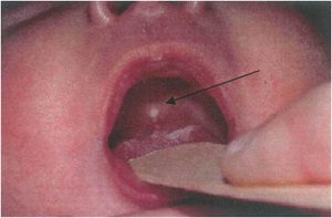
Bohn nodule
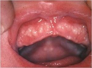
Whitish-yellow nodules which appear on the gums and hard palate in a large percentage of new-born babies.
Causes
Bohn’s nodules are odontogenic cysts that arise from the dental lamina. They are filled with keratin.
Epstein pearls are epithelial inclusion cysts.
The condition is harmless but worries new mothers who may mistake the nodules for emerging teeth.
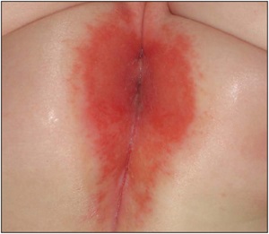
Perianal dermatitis of neonate
is an area of erythema centred on the anus and occasionally accompanied by erosion and bleeding.
Early lesions are small erythematous macules 2 – 3 mm in size.
This condition is usually attributed to formula milk feeding in the newborn and abnormal faecal pH .
It is observed between the fourth and seventh days of Life .
Perianal dermatitis is 6.5 times more frequent in premature babies.
Cradle cap
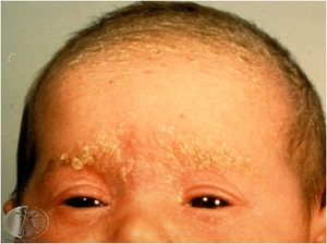
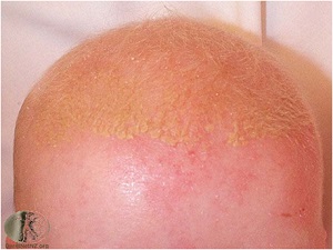
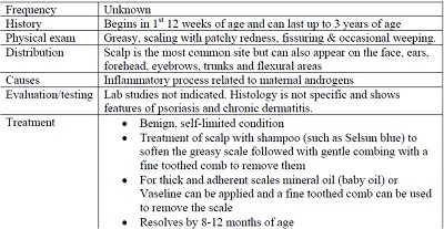
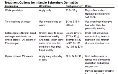
Seborrheic dermatitis is an extremely common rash characterized by erythema and greasy scales Many parents know this rash as “cradle cap” because it occurs most commonly on the scalp.
Other affected areas may include the face, ears, and neck. Erythema tends to predominate in the flexural folds and intertriginous areas, whereas scaling predominates on the scalp.
Because seborrheic dermatitis often spreads to the diaper area, it is important to consider in the evaluation of diaper dermatitis

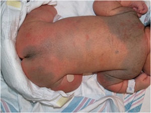
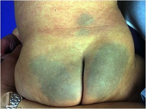
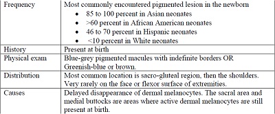
Mongolian spot
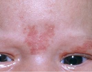
Stork bite
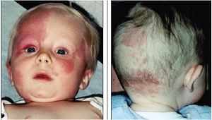
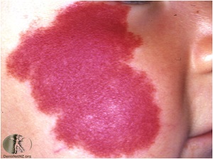
Port wine stain
Angel kiss

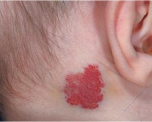
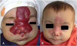
Infantile hemangioma
آقاي ساسان سلام
اگر بيماري در حال پيشرفت باشد،نقاط جديد بدن كه تازه دارند رنگدانه از دست مي دهند ولي هنوز روند پيشرفت ادامه دارد،خيلي سفيد نيستند،در خصوص سوال دوم،چنين آمپول و درماني وجود ندارد،بهترين درمان براي شما نور درماني هست .
زمان بهترین و ارزشمندترین هدیه ای است كه می توان به كسی ارزانی داشت.هنگامی كه برای كسی وقت می گذاریم، قسمتی از زندگی خود را به او میدهیم كه باز پس گرفته نمی شود . باعث خوشحالی و افتخار من است كه برای عزیزی مثل شما وقت می گذارم و امیدوارم كه با راهنماییهای اساتید این رشته واظهار نظر شما عزیزان این سایت آموزشی پر بارتر گردد.