آقاي ساسان سلام
اگر بيماري در حال پيشرفت باشد،نقاط جديد بدن كه تازه دارند رنگدانه از دست مي دهند ولي هنوز روند پيشرفت ادامه دارد،خيلي سفيد نيستند،در خصوص سوال دوم،چنين آمپول و درماني وجود ندارد،بهترين درمان براي شما نور درماني هست .
Dermatoscopic examination of hair and scalp is known as trichoscopy.
Here, we summarize the literature on the dermatoscopic diagnosis of congenital and acquired hair shaft disorders.
As hair shaft disorders are rare, most published articles on their dermatoscopic features are limited to case reports or series.
Dermatoscopy is fast, noninvasive, and cost efficient and allows for easy office diagnosis of all hair shaft abnormalities .
Conditions such as pili trianguli and canaliculi that are not recognizable by examining hair shafts under the light microscope.
Dermatoscopy is fast, noninvasive, and cost efficient and allows for easy office diagnosis of all hair shaft abnormalities .
Conditions such as pili trianguli and canaliculi that are not recognizable by examining hair shafts under the light microscope.
Most hair shaft abnormalities in fact do not affect the entire scalp but are limited to a few hairs and it can be difficult to detect the abnormality in a random sample.
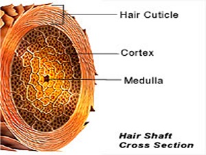
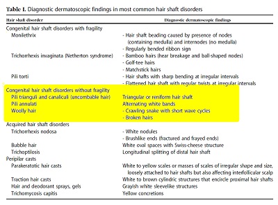
HAIR SHAFT ABNORMALITIES
congenital
acquired
Congenital disorders can be an isolated finding or can be a part of complex syndromes.
Some abnormalities are associated with structural weakness of the shaft and, therefore, with hair breakage and loss.
Increased fragility and breakage is the main feature that allows classification of these disorders.
Hair shaft disorders with increased fragility can be:
congenital
monilethrix
pili torti
trichorrhexis invaginata [TI]
Trichothiodystrophy
trichorrhexis nodosa [TN]) .
acquired (TN, bubble hair).
Acquired disorders are more common than congenital.
Hair shaft disorders without increased fragility are only congenital and include:
pili annulati,
woolly hair,
Uncombable hair syndrome,
pili bifurcati ,
multigemini.
NORMAL HAIR SHAFTS
Normal hair shafts are uniform in shape and color with continuous interrupted, fragmented, or absent medulla. The hair fibers are straight and parallel to the long axis of the hair.
CONGENITAL HAIR DISORDERS WITH INCREASED FRAGILITY
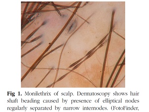
Monilethrix
Monilethrix is a hair shaft disorder characterized by hair fragility and alopecia as a result of hair breakage.
It is an autosomal dominant disorder caused by mutations of the human basic hair keratins hHb6 and hHb1, which most likely cause disruption of the Keratin formation in the hair shaft cortex.
As the hair shaft in monilethrix is easily broken, the clinical presentation is of hypotrichosis most pronounced on the occipital scalp, which is more exposed to friction.
The severity of monilethrix considerably varies even among members of the same family and may range from almost normal scalp to total alopecia.
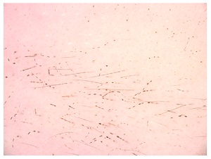
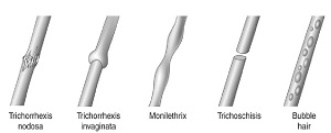
Dermatoscopy shows hairs haft Beading caused by the presence of elliptical nodes, regularly separated by narrow internodes that are the site of fracture (dystrophic constrictions).
The nodes have a diameter of a normal hair and a medulla, whereas the internodes have no medulla.
Rakowska et al coined the term ‘‘regularly bended ribbon sign’’ in monilethrix as the hairs with beaded appearance bent regularly at multiple locations with a tendency to curve in different directions.
This together with the absence of medulla differentiates monilethrix from pseudomonilethrix.
The extent of hair breakage in monilethrix is variable and can easily be assessed with Dermatoscopye in severely affected patients vellus hairs also show the abnormality.
Eyebrows can also be affected .
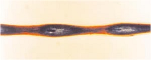
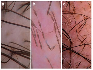
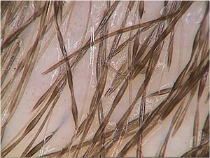
TI (bamboo hair)
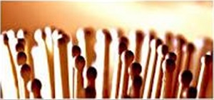
On light microscopy the hair shaft in TI shows multiple knots, resembling the ball-in-cup rings Of bamboo.
These are caused by invagination of the distal portion of the hair shaft into its Proximal portion.
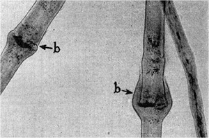
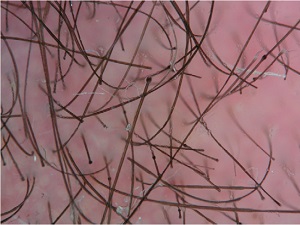
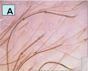
TI may be isolated, but most commonly is part of the Netherton syndrome, a rare autosomal recessive genodermatosis that combines ichthyosis,bamboo hair, and atopic dermatitis.
Clinically the hair is sparse, brittle, and short.
The diagnosis of Netherton syndrome may be established on the basis of just 1 abnormal hair.
However, because the abnormalities may be limited to a few hairs, especially in adults, it is often difficult to find a hair with Pathognomonic features in the sample submitted for light microscopic examination.
Plucking hairs is often not helpful either, because the most Abnormal and diagnostically useful hairs are short and forceps miss them
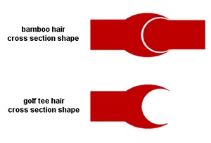
Dermatoscopy has been described as hair breakage with ball-shaped nodes as well as invaginations and golf-tee
endings .
This feature is observed also in the eyebrows, which may be the sole site of involvement in many patients with Netherton syndrome and more prominent and easier for the diagnosis than the scalp
Goujon et al described 3 types of dermatoscopic features in the eyebrows in Netherton syndrome:
bamboo hairs, golf-tee hairs, and matchstick hairs, which present as short hair shafts with a bulging tip most likely
Caused by breakage of the bamboo hairs.
The same 3 findings have been reported also in the Eyelashes in Netherton syndrome.
The golf tee-like ends are better visualized at higher magnification (*70).
Pili torti
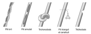
Pili torti are flattened hairs and present twisting of the hair shaft through 180 degrees at irregular intervals.
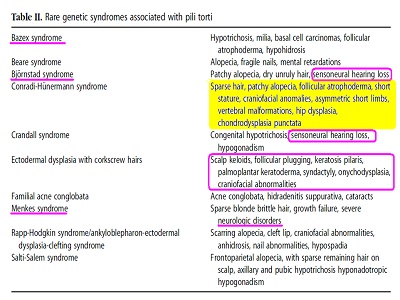
Pili torti are common in patients with other congenital hair shaft disorders and are a feature of several rare genetic syndromes.
They can also be observed in the normal scalp and in Cicatricial alopecias (particularly lichen planopilaris, Frontal fibrosing alopecia, and central centrifugal Cicatricial alopecia).
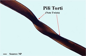
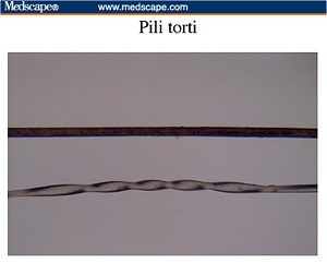
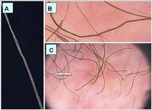
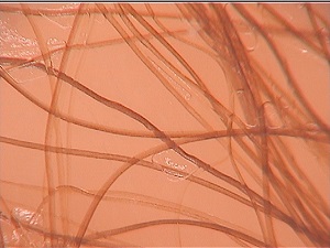
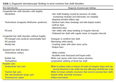
Pili torti. Dermatoscopy shows hair shaft with sharp bending at irregular intervals.
A recent dermatoscopic study found pili torti in approximately 57.1% of cases of primary cicatricial alopecia and considered them another clue to the diagnosis.
At low magnification (*20) dermatoscopy Demonstrates hair shafts with sharp bending at Irregular intervals.
At higher magnifications pili torti show flattened hair shafts with regular twists at Irregular intervals.
Trichothiodystrophy
Trichothiodystrophy’’ is a term that describes cystine-deficient brittle hair.
The characteristic tiger-tail pattern is not visible On Dermatoscopy and can be viewed with Polarized microscopy only.
According to Rakowska et al dermatoscopy of The hair shaft in trichothiodystrophy is nonspecific:
the affected shaft shows nonhomogenous structure of ‘‘grains of sand’’ and slightly wavy contour.
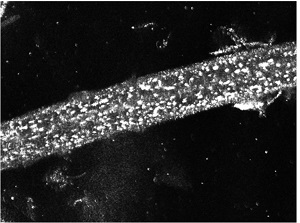
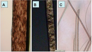
Congenital TN
TN can rarely be congenital.
Congenital TN is reported in argininosuccinic aciduria and citrullinemia, which is an inborn error of the urea synthesis.
It may also occur as an isolated clinical abnormality in sporadic case, or in families with abnormal fragility of the hair becoming evident shortly after birth
Congenital TN has been described associated with 20-nail dystrophy.
It has also been reported as part of Menkes kinky hair syndrome (Menkes disease) together with monilethrix and pili torti. As in other hair shaft disorders, hair breakage mostly affects scalp areas exposed to friction and dermatoscopy allows for rapid differentiation from more common disorders such as monilethrix.
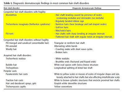
HAIR SHAFT DISORDERS WITHOUT INCREASED FRAGILITY
Pili trianguli and canaliculi (uncombable hair)
Pili trianguli and canaliculi present as uncombable,dry, Unruly hair.
The condition affects children and improves Spontaneously with aging.
It can not be diagnosed under the microscope and Scanning electron microscopy is considered the gold standard for the diagnosis.
uncombable hair
Recently dermatoscopy was shown to be very useful for this diagnosis and it shows triangular or reniform hair shaft
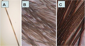
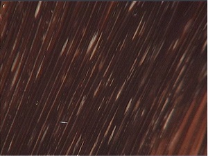
with Longitudinal groove or flattening.
Pili annulati
Pili annulati is an autosomal dominant hair shaft disorder characterized by alternating light and dark bands in hairs of affected individuals.
The pathogenesis is not known but the abnormality is caused by the presence of air filled cavities within the hair shaft.
At dermatoscopy the hair shaft shows alternating white bands that correspond to the air-filled cavities .
This differs from what is seen under Light microscopy where the cavities appear darker than the cortex.
Intermittent medulla observed in individuals with thick hair shafts can be misdiagnosed as pili annulati.
Pili annulati are more susceptible to weathering Than normal hair and TN is frequently detected in the hair shafts affected by the abnormality.
Association of pili annulati with alopecia areata has been reported.
Woolly hair
Woolly hairs are shaft anomalies that clinically present as tightly curled hair.
dermatoscopy can distinguish healthy curly hair from woolly hair syndrome.
In woolly hair dermatoscopy shows the image of hair shafts resembling a crawling snake with short wave cycles.
Apart from that, trichoscopy also demonstrates Broken hair shafts, which correspond to increased fragility of hair shafts caused by longitudinal twisting.
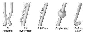
Pili bifurcati and multigemini
In pili bifurcati and multigemini, the hair shafts grow from the same papilla that splits from a single tip to a double tip in pili bifurcati and to Several Splits in pili multigemini.
Irregularities in configuration of the hairs, longitudinal grooving, and areas of bifurcation and re-adhesion of the hair shafts are common.
Pili bifurcati and multigemini
Pili bifurcati is uncommon and has been reported in normal hair and associated with pili canaliculi And monilethrix.
No reports exist on the dermatoscopic features in these conditions.
ACQUIRED HAIR SHAFT DISORDERS
Trichorrhexis nodosa
TN is an acquired hair shaft disorder, which results from hair weathering.
It mainly affects the free ends of long hair(distalTN).
Causes of TN include mechanical, chemical, and thermal damage, mainly as a result of hairstyling and processing.
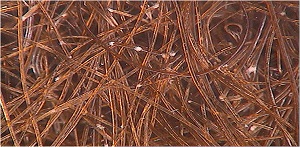
The white nodules are easily seen on Dermatoscopy that shows deterioration of the hair shaft in correspondence with the nodules.
High magnification shows these fibers in detail, whereas at low magnification they look as light colored nodules or gaps located along the hair Shaft.
Fractured and frayed ends have a brushlike appearance on dermatoscopy.
Dermatoscopic examination also allows for diagnosing hair breakage caused by TN in the shed broken hair shaft.
This can be useful to quickly diagnose acute patchy alopecia caused by proximal TN after hair perming or straightening.
TN can rarely be a result of systemic causes Including metabolic disorders or malnutrition.
Donati et al described TN in a child with Phenylketonuria.
Bubble hair
Bubble hair occurs when bubbles form within the hair cortex as a result of high temperatures from styling with blow dryers or curling irons.
Bubble originates within the hair because of gas Formation when the wet hair shaft is rapidly exposed to excessive temperatures.
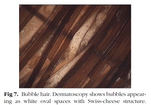
At dermatoscopy bubbles appear as white oval spaces with Swiss-cheese structure as they Contain Gas.
Trichoptilosis
Trichoptilosis (split ends) is longitudinal splitting of the distal end of the hair.
It is the most common sign of weathering; when seen in short hairs it can be caused by friction or trichotillomania.
Dermatoscopy shows longitudinal splitting of the distal hair shafts. Splitting produces 2 or multiple frayed ends of different length.
PERIPILAR CASTS
This group includes hair disorders associated with infestations of the scalp with fungi (tinea capitis, trichomycosis capitis), bacteria, pediculosis, and deposits from paints, glue, and hair spray.
Piedra (trichosporosis)
In white piedra the hair shafts appear coated by yellow or beige sheaths.
The nodules Measure 1 to 1.5 mm in diameter;
they are fusiform and of soft consistency, mainly attached to the distal portions of the hair.
In black peidra dark pebble-sized nodules are distributed irregularly along the length of the Hair shaft of the scalp, eyebrows, eyelashes, or Beard.
Trichomycoses affect usually axillary and pubic/ scrotal hairs and therefore this location on the scalp is unusual.
However, in this case maceration and constant lying in bed because of cerebral palsy were considered the cause of corynebacterium colonization of the hair shafts.
Dermatoscopy identified yellow concretions attached to the hair shafts.
Hair casts
Differential diagnosis among peripilar casts (scarring alopecia), interfollicular scaling (psoriasis, Seborrheic dermatitis), or nits (pediculosis) and pseudonits (hair sprays) may be very difficult when assessed only by the naked eye.
Dermatoscopy is an easy method to identify the correct type of cast.
Hair casts are a sign of ongoing traction in patients with traction alopecia.
On dermatoscopy traction hair casts appear as white to brown cylindric Structures with spindled edges that encircle the Proximal hair shafts.
Dermatoscopy enables differentiating hair casts from nits in pediculosis and from pseudonits caused by hair sprays and gels.
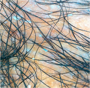
Traction alopecia. Dermatoscopy shows cylindric white hair casts encircling several hair shafts.
CONCLUSION
Trichoscopy is a fast and noninvasive new tool in the diagnosis of hair shaft abnormalities.
Handheld dermatoscopes allow for magnification of *10 to *20 and videodermatoscopes allow for Magnifications up to *160.
Both types of devices enable the dermatologists to distinguish fractures, irregularities, twisting, and extraneous matter without plucking or cutting hairs for microscopic examination.
yellow dots, exclamation point hairs and dystrophic hairs are characteristic features; the color of the yellow dots may vary from yellow to yellow-pink. Note some vellus-like hair shafts.
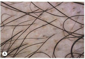
Androgenetic alopecia (AGA):a diversity of hair shaft diameters affects >20% of the hairs. Brown halos around hair shafts (peripilar sign) is a subtle clue.
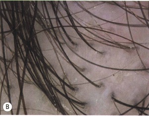
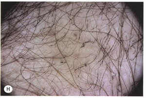
Telogen effluvium: the hair shafts are of equal diameter (unlike AGA).
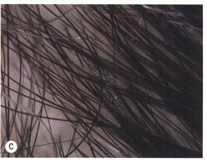
Tinea capitis: comma hairs are a clue to the diagnosis.
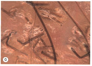
Monilethrix: the presence of broken hair shafts plus hairs with a beaded appearance allows a bedside diagnosis.
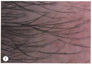
Trichorrhexis invaginata (bamboo hairs) in a patient with Netherton syndrome: broken hair shafts and nodose swellings.
آقاي ساسان سلام
اگر بيماري در حال پيشرفت باشد،نقاط جديد بدن كه تازه دارند رنگدانه از دست مي دهند ولي هنوز روند پيشرفت ادامه دارد،خيلي سفيد نيستند،در خصوص سوال دوم،چنين آمپول و درماني وجود ندارد،بهترين درمان براي شما نور درماني هست .
زمان بهترین و ارزشمندترین هدیه ای است كه می توان به كسی ارزانی داشت.هنگامی كه برای كسی وقت می گذاریم، قسمتی از زندگی خود را به او میدهیم كه باز پس گرفته نمی شود . باعث خوشحالی و افتخار من است كه برای عزیزی مثل شما وقت می گذارم و امیدوارم كه با راهنماییهای اساتید این رشته واظهار نظر شما عزیزان این سایت آموزشی پر بارتر گردد.